- OCT
OCT Principle
We present a brief overview of the principle of OCT to aid in your product selection.
Tutorial 1
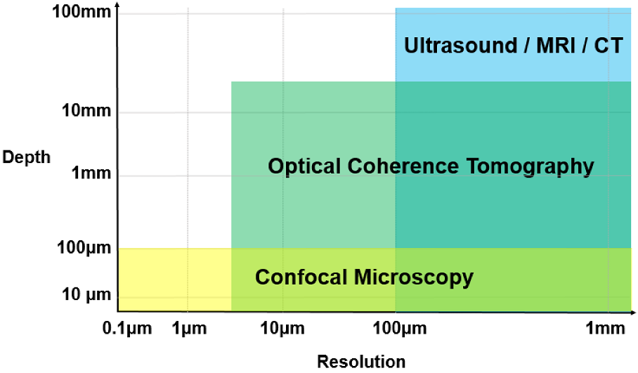
Optical Coherence Tomography (OCT) is a non-invasive imaging technique that provides subsurface imaging of tissue or other materials. OCT is an interferometric imaging modality that combines the coherent backscattered light from a sample with a reference taken directly from the light source. OCT based diagnostic systems are now widely used in ophthalmology. Compared to other medical imaging technologies such as Magnetic Resonance Imaging (MRI), Positron Emission Tomography (PET), X-Ray Computerized Tomography (CT) and Ultrasonography, OCT provides a safe, high resolution solution at a cost that enables widespread use in hospitals and clinics.
There are a number of distinct types of OCT. Early OCT systems were based on time-domain optical interferometry in which the optical path length difference between the reference and sample was modulated with time. Time-Domain (TD) OCT demonstrated many potential applications but scan speed and other performance limitations has limited its commercialisation. Most commercial systems now use a Fourier-Domain (FD) approach in which the wavelength of light is varied and depth information is extracted by taking the Fourier transform of the detected interferometric signal.
Tutorial 2
Fourier Domain (FD) OCT works by analyzing the individual frequency components of backscattered light from the sample or tissue. There are two distinct FD-OCT modalities. In Spectral-Domain OCT (SD-OCT) a low coherence light source is used in together with a spectrometer. Frequency components are detected spatially on the spectrometers CCD array. Fast CCD sensors provide for fast imaging together with a signal-to-noise ratio (SNR) that is 20-30 dB better than TD-OCT. However, there are a number of disadvantages. Any motion of the sample during imaging leads to washout of the interference fringes. This occurs when the interference pattern moves over multiple CCD pixels within the integration time. Further, the development of higher performance SD-OCT has been limited by the availability of high speed, high resolution InGaAs CCD sensors.
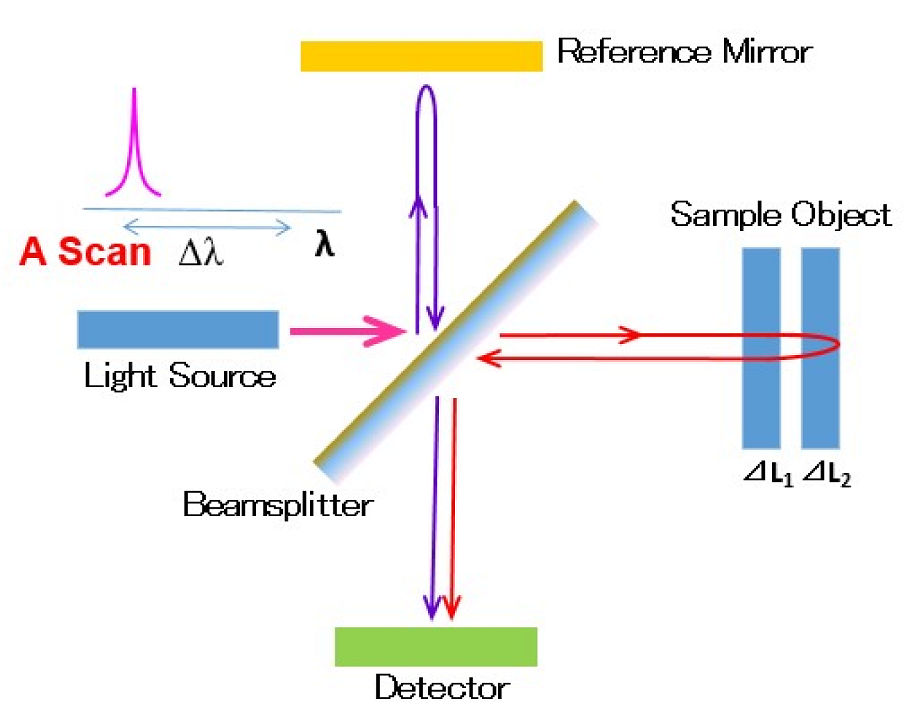
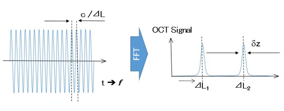
In Swept-Source OCT (SS-OCT) the spectral domain broadband source is replaced by a laser that rapidly and repeatedly sweeps the wavelength over a broad range of wavelengths, and the spectrometer is replaced by a single photo-detector. As the laser sweeps, the interferometric signal is detected sequentially by the photodetector. A single sweep corresponds to an A-scan. Reflections are detected simultaneously from all depths and by performing a Fourier transform on the detected interferogram this is converted to a depth dependent reflection profile. Repeating the A-scan at different points on the sample produce a two dimensional cross section or B-scan. This technique achieves the same sensitivity benefit as SD-OCT while eliminating the disadvantage of fringe washout. It is also more flexible as reasonably priced light-sources are available at longer wavelengths (>1.0 µm) and with extremely high scanning rates.
Wavelength Range
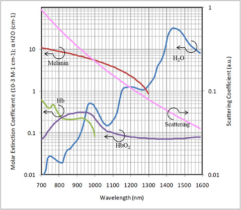
OCT systems operate at a variety of different wavelengths. The choice of the wavelength will depend on the transparency at that wavelength together with the water absorption and scattering properties of the material of interest. 800 nm has been widely used for retinal imaging because of the low absorption in the vitreous humor. More recently, 1060 nm is preferred for this application because of the large penetration in retinal tissue as well as the low dispersion. For the endoscopic applications, wavelength of 1310 nm or wider are used because of the low-scattering, which result the deeper depth penetration in tissue. The 1310 nm range is also preferred for many applications because of the lower cost and wider availability of optical components that are available because of the synergy with fiber optics in telecommunications.
Speed & Range
Imaging speed corresponds directly to the scanning speed, or repetition rate of the wavelength sweep of the swept-source laser. In SS-OCT the scanning speed corresponds to the A-line rate. Increasing the A-line rate makes it possible to accommodate more A-lines per frame or increase the frame rate. In practice, a fast A-line rate removes imaging artifacts associated with sample movement and enables the rapid imaging a large area/volume measurement without compromising resolution. Typically, a scanning speed up to 400 kHz is used.

Imaging depth is mainly limited by the penetration depth of the light in the sample. It is also limited by the number of data points and the coherence length of the light source. As indicated in the Nyquist Theorem for FD-OCT, a shorter sampling interval provides a longer (deeper) imaging depth. In SS-OCT, this corresponds to the sampling speed of the data acquisition card and the scanning speed of light source. On the other hand, for higher frequency components the signal intensity drops, i.e. at a deeper range. The OCT signal is a convolution of the light source spectrum and the interferogram. Since the instantaneous linewidth is finite a longer coherence length gives lower signal intensity drops. Coherence length is defined as the optical round trip delay or twice of the depth range where fringe visibility drops by half or the Fourier-transformed OCT signal drops by 6 dB compared to the signal power at zero delay.

Resolution
In SS-OCT axial (depth) resolution is related to the wavelength range as well as the center wavelength of the laser. A wider scanning range corresponds to a higher axial resolution. For the same scan range, a shorter centre wavelength also provides a higher axial resolution.It should be noted, though, that when selecting the centre wavelength the absorption and scatting properties of the sample must also be taken into consideration.
The lateral resolution is related to the NA (numerical aperture) of the lens used in the probe head. A higher NA gives higher lateral resolution at the beam waist depth position, but a higher NA limits the depth of focus. When determining the lens used for imaging, consideration must therefore be made of both the required resolution and depth of focus. Santec IVS systems support a number of lens configurations to optimize these parameters for different applications.

